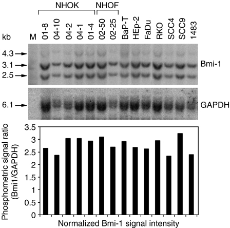
Figure 2.
Bmi-1 gene is not amplified in OSCC. Genomic DNAs from five different NHOK strains, two NHOF strains, and seven cancer cell lines were digested with EcoR I and Hind III and transferred for probing. Radiolabelled probes synthesised from Bmi-1 or GAPDH cDNA were hybridised sequentially onto the membrane. The phosphometric intensities were plotted as the ratio of Bmi-1 to GAPDH. The lack of statistical difference (P>0.05) in the levels of Bmi-1 radioactive signals was determined by unpaired T-test (one-ways ANOVA) between the NHOK and the cancer groups.

