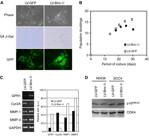
Figure 4.
Inhibition of endogenous Bmi-1 causes premature senescence in NHOK. (A) Rapidly proliferating NHOK (05-10) was infected with LV-GFP or LV-Bmi-1i. Phase contrast photographs, SA β-Gal staining, and GFP fluorescence were obtained 10 days after virus infection. Original magnification, × 100. (B) Proliferation kinetics of NHOK infected with LV-GFP or LV-Bmi-1i was determined and plotted against time in culture. (C) NHOK cultures were harvested at 10 days after infection with LV-GFP or LV-Bmi-1i, and the expression levels of GPR1, Cyc2A, MMP-1, and MMP-3 were determined by semi-quantitative RT–PCR. The band intensities were quantitated and plotted by Scion Image software against those of GAPDH amplification. (D) NHOK (05-10) and SCC4 cells infected with LV-GFP or LV-Bmi-1i were harvested at 10 days after infection, and Western blotting was performed with 100 μg WCE for p16INK4A. CDK4 was detected as a loading control.

