Figure 5.
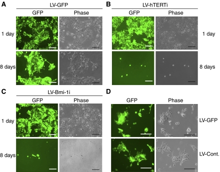
Inhibition of endogenous Bmi-1 led to effective loss of viability in BaP-T cells. Rapidly proliferating BaP-T cells were infected with LV-GFP (A), LV-hTERTi (B), LV-Bmi-1i (C), or LV-Cont (D). (A–C) The infected cells were labelled with green fluorescence owing to the GFP expression from the pLL3.7 parental lentiviral vector and shown along with the phase contrast view after 1 day or 8 days post-infection. The cells infected with LV-GFP were passaged owing to reaching confluence after 3–4 days, whereas those infected with LV-Bmi-1i or LV-hTERTi were maintained in the same dish without passaging for the period of observation. (D) In another experiment, BaP-T cells were infected with LV-GFP or LV-Cont. and maintained in parallel for 9 days and photographed. In this experiment, no notable differences in cellular morphology, replication capacity, or viability were noted between the two groups. Bar=200 μm.

