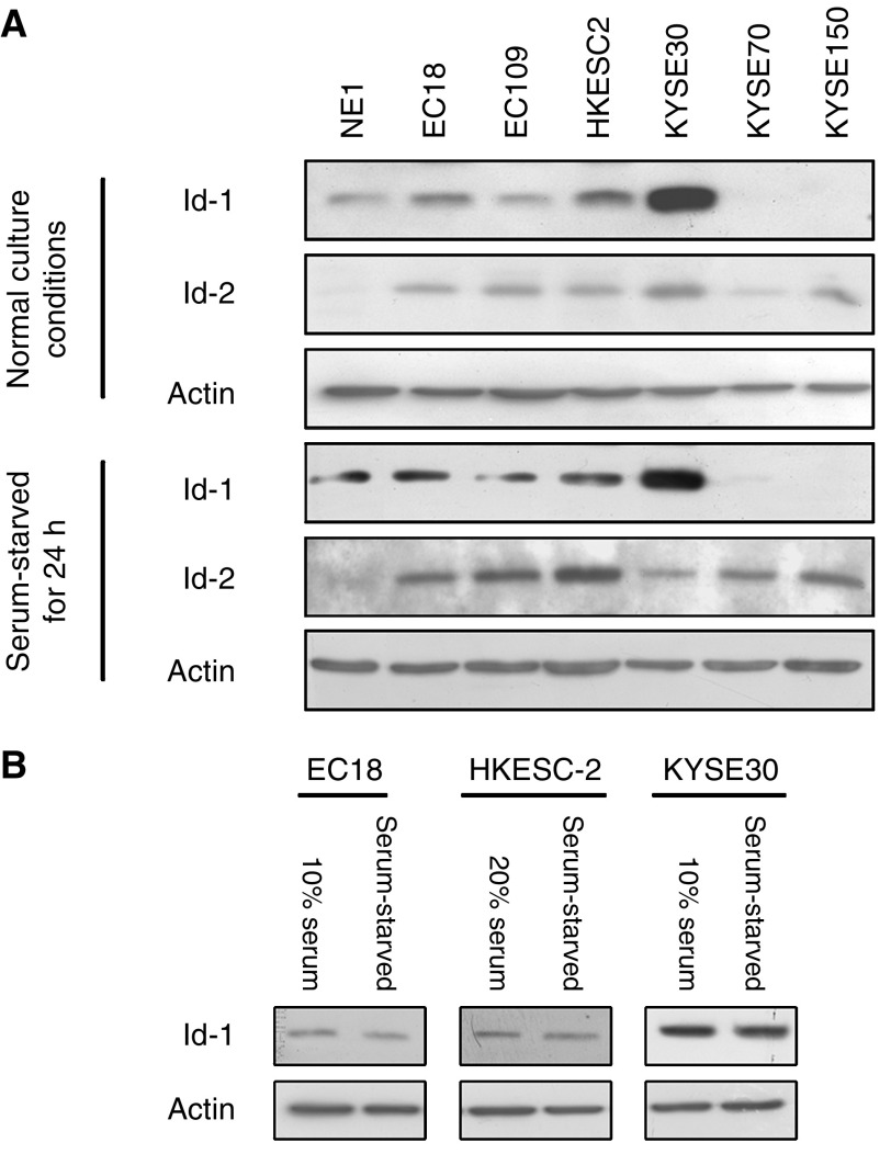
Figure 1.
Western blot analysis of Id-1 and Id-2 expression in immortalized oesophageal epithelial and ESCC cells. Cancer cells (2 × 10⁵) were seeded in six-well plate with supplement of serum, according to the recommendation from the suppliers of the cell lines, as described in Materials and Methods. Medium was changed after 1 day with either serum-free medium or fresh serum-supplemented medium NE1 was cultured in KSFM with growth factors supplementation, and fresh medium was replaced after 1 day. Cells were harvested after 24 h. Protein was extracted and the expression level of Id-1 and Id-2 was then compared by Western blot analysis. Note that the expression of Id-1 was detected in four out of six ESCC cell lines while Id-2 was expressed in all ESCC cell lines tested. (B) The expression level of Id-1 in three ESCC cancer cell lines with or without serum supplementation. Id-1 expression level was slightly reduced when the cells were serum-starved for 24 h.

