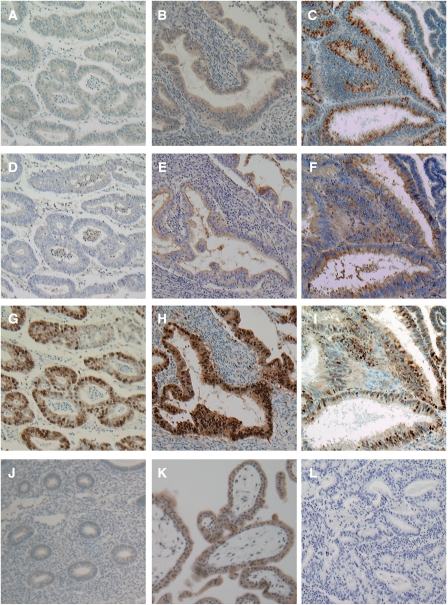
Figure 2.
Immunohistochemical staining patterns for GnT-V and staining of L4-PHA in endometrial cancers. Staining pattern of a tumour: (A) GnT-V low; (B and C) GnT-V high. (D–F) L4-PHA staining and (G–I) PCNA immunostaining were performed simultaneously with the same A, B, and C specimens, respectively. (J) Normal endometrial cells showed very faint or negative GnT-V expression. (K) Positive control for GnT-V (normal placenta). (L) Negative control. Original magnification, × 100.

