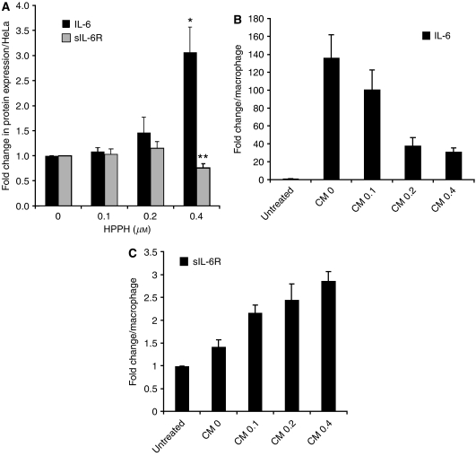
Figure 1.
Induction of IL-6 and sIL-6Rα following PDT. (A) HeLa cells were treated with increasing concentrations of HPPH and irradiated with 1 J cm−2 light. Conditioned media were collected at 24 h post PDT. (B, C) Pulmonary macrophages were incubated for 24 h with CM from PDT-treated HeLa cells. The concentrations of IL-6 and sIL-6Rα in the supernatant culture media were determined by Bio-Plex/Luminex and ELISA, respectively. The values in each experimental series were normalised to the untreated controls and the relative changes determined in three independent experiments were expressed as mean and SD. Values indicated by stars denote difference with P<0.05 compared to controls. As reference, the average concentrations for control cultures were as follows: (A) IL-6: 180 pg ml−1, sIL-6Rα: 10 pg ml−1; (B) IL-6 for CM-0 culture: 32 ng ml−1; (C) sIL6Rα: 100 pg ml−1.

