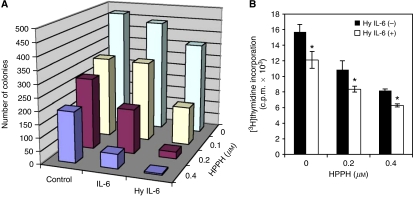
Figure 3.
Interleukin-6 and Hyper-IL-6 suppress proliferation of PDT-treated HeLa cells. (A) HeLa cells were treated with increasing concentrations of HPPH and exposed to 1 J cm−2 of light. After irradiation, cells were plated in 6 cm dishes in the presence or absence of IL-6 cytokines (IL-6 100 ng ml−1; Hyper-IL-6 160 ng ml−1). After 14 days, colonies were fixed, stained with crystal violet and counted (colonies with >25 cells). The values represent means of a single experiment performed in triplicate, representative of three performed. (B) [3H]thymidine incorporation into HeLa cells. Post –PDT, cells were treated or not treated with 160 ng ml−1 Hyper-IL-6. [3H]Thymidine incorporation after 48 h of treatment is plotted as the mean±s.d. of a single experiment performed in triplicate, representative of three independent experiments. *P<0.05, compared with the corresponding value of PDT alone.

