Figure 2.
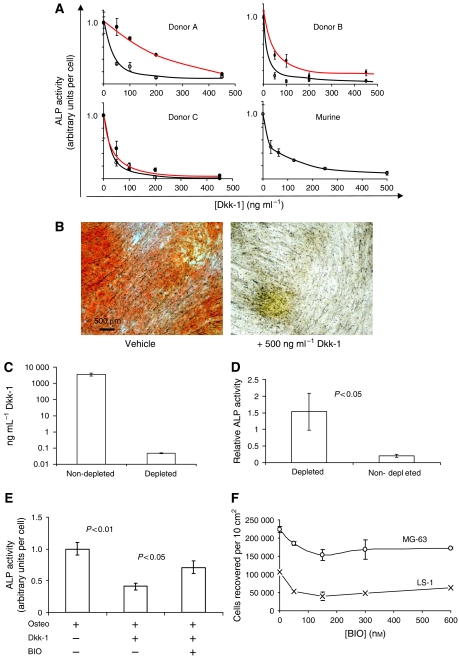
(A) Osteogenic differentiation of MSCs in the presence of Dkk-1. Results from cells derived from three human donors and pooled murine donors are presented. Osteogenic differentiation is presented as a function of membrane ALP activity, an early marker of osteogenesis. Measurements are normalised to control levels of activity, designated 1.0. The black lines represent MSCs prepared from the fluid component of bone marrow, and the red lines represent MSCs prepared from bone spicules filtered from the aspirates. Dkk-1 exposure causes a dose-dependent inhibition of alkaline phosphatase activity. (B) Alizarin Red stained, long-term cultures of osteogenic MSCs in the presence and absence of Dkk-1. Calcium detection by Alizarin Red S demonstrates that Dkk-1 inhibits mineralisation of the cultures. (C) Immunodepletion of Dkk-1 from MG63 OS conditioned medium through incubation with a polyclonal antibody against Dkk-1. The Dkk-1–antibody complexes were removed from the medium by protein A affinity chromatography, then the medium was assayed by ELISA. (D) Osteogenic differentiation by MSCs in the presence of nondepleted and Dkk-1 immunodepleted conditioned medium from MG63 OS cells. Representative results from one out of three donors are presented. Measurements were achieved by ALP assay, values represent the mean (n=6), and error bars represent s.d. P-values were calculated by two-tailed Student's t-test. (E) Osteogenic differentiation by MSCs in the presence of Dkk-1 and with or without the GSK3β inhibitor BIO. (F) The effect of a range of BIO doses on the proliferation of OS cells. Cell numbers were evaluated by fluorescent nucleic acid intercalation assay.

