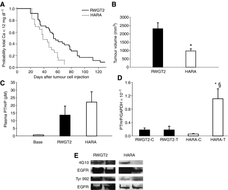
Figure 4.
Comparison of RWGT2 and HARA models of hypercalcaemia. (A) Kaplan–Meir analysis of time to the development of hypercalcaemia. Fifty percent of mice developed HHM 40 days after injection of HARA cells compared to the RWGT2 xenograft mice that developed HHM 60 days after injection of cells. P=0.01. RWGT2 tumours (n=18) and HARA tumours (n=10). (B) Average tumour volume at time of hypercalcaemia. When hypercalcaemia was diagnosed, the RWGT2 (n=18) tumours were nearly twice as large as the HARA tumours (n=10) *P=0.04 HARA compared to RWGT2. Two-tailed Student's t-test. (C) Comparison of plasma PTHrP concentrations in hypercalcaemic untreated mice with RWGT2 and HARA xenografts. Plasma PTHrP concentrations were determined 78 h after the development of HHM in both xenograft models. Average plasma PTHrP concentrations in the untreated mice were 80% higher (22 vs 12 pM) in the HARA (n=5) as compared to RWGT2-bearing mice (n=8). (D) Comparison of PTHrP mRNA expression between cells grown in vitro and in vivo. RNA was extracted from tumours that were removed 78 h after hypercalcaemia was identified. The ratio of PTHrP to GAPDH mRNA was assayed by QRT-PCR. The ratio of PTHrP to GAPDH mRNA in HARA tumours (HARA-T) was increased 100-fold compared to HARA cells (HARA-C) grown in vitro. The ratio of PTHrP to GAPDH mRNA in HARA tumours was sixfold higher than RWGT2 tumours (RWGT2-T). Values in all panels represent the mean of four samples from individual cultures or tumours±s.e.m. *P=0.027 HARA tumours relative to HARA cells; §P<0.05 HARA tumours relative to RWGT2 tumours. The QRT-PCR was repeated twice with similar results. Two-tailed Student's t-test. (E) Phosphorylation of the EGFR was measured in protein extracts from RWGT2 and HARA tumours by Western blotting. The general phosphotyrosine antibody 4G10 and polyclonal antibody for phosphorylated Tyr 992 was used to probe 1 mg of extracted tumour protein precipitated with conA-Sepharose. Blots were stripped and reprobed with EGFR antibodies. Four tumours from each cell line were evaluated.

