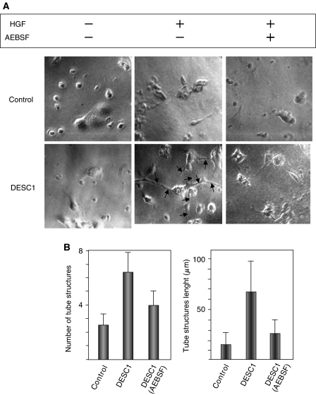
Figure 4.
(A) HGF-stimulated MDCK/DESC1 cells embebbed in collagen gel form tubular structures. MDCK cell clones stably transfected with control vector or expressing DESC1 were cultured in a type I collagen matrix for 7 days in presence (+) or in absence (−) of 30 ng ml−1 of HGF, and in the presence (+) or absence (−) of 50 μM of AEBSF. The branching extensions formed by MDCK/DESC1 cells are indicated by arrows. (B) Quantification of the number and length of tubular structures of 15 randomly selected microscopic fields following HGF stimulation.

