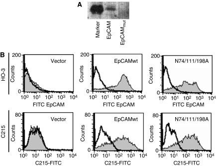
Figure 3.
HO-3 and C215 recognise EpCAM independently of the glycosylation status. (A) HEK293 cells were stably transfected with an expression vector for wild-type EpCAM and a triple mutant lacking all glycosylation sites (EpCAMmut). Equal amounts of cell lysates of each cell lines were separated by 10% SDS–PAGE and subjected to glycostaining. Staining was assessed with the FLA 5000 scanning device (Fuji). M represents an internal marker provided by the manufacturers. (B) HEK293 cells stably expressing wild-type EpCAM, a triple mutant lacking all N-glycosylation consensus sites (N74/111/198A; EpCAMmut), or the empty vector for a control were stained with HO-3 (grey curves; upper panels) or C215 (grey curves; lower panels) in combination with a FITC-conjugated secondary antibody. As a control, primary antibody was omitted (black line). All data are representative results from three independent experiments.

