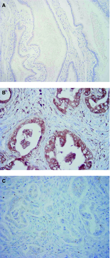
Figure 1.
Immunohistochemical staining for MK in invasive ductal adenocarcinoma of the pancreas head. (A) Normal pancreatic ductal epithelium dissected approximately 3 cm apart from the cancerous region (× 200). Note that almost all cells are unstained or stained very slightly by the antibody. (B) Carcinoma cells positively stained by MK antibody (× 400). Note cytoplasmic staining for MK. (C) Carcinoma cells stained negatively by MK antibody (× 400).

