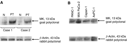
Figure 2.
Western blotting for MK protein. (A) Pancreatic primary tumours (PT) and non-cancerous regions (N) dissected approximately 3 cm apart from the cancerous region. Cases 1 and 2 are from each independent patient. (B) Pancreatic carcinoma cell lines. Note the presence of a 13-kDa band corresponding to MK protein in primary tumours and cultured cells. The presence of a 43-kDa band corresponding to β-actin protein in each sample indicates that samples are equally loaded.

