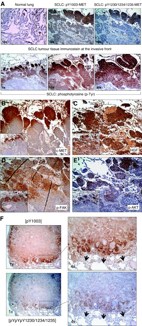
Figure 5.
Topographic analysis of the invasive front of SCLC using phosphoantibody IHC. (A) Topographic role of p-MET and phosphoproteins with pTyr activation. (B) Overexpression of c-MET along the SCLC invasive tumour front, × 10. Inset: × 4. (C) Overexpression of HGF in SCLC tumour tissue, × 10. Inset: × 4. (D) Topographic role of activated cytoskeletal focal adhesion protein p-FAK, × 10. Inset: × 4. (E) Topographic role of activated survival signalling molecule p-AKT, × 10. Inset: × 4. (F) Preferential expression of activated p-MET along the tumour invasive front in lung adenocarcinoma. Upper panel, p-MET [Y1003]. Lower panel, p-MET [pY1230/1234/1235]. Magnification: left, × 1; right, × 4.

