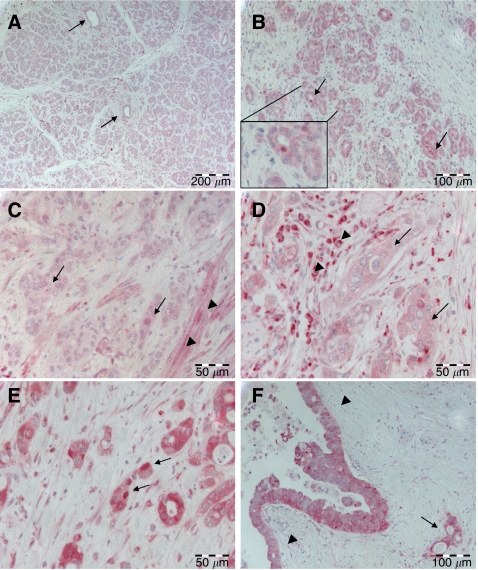
Figure 1.
RelA/p65 expression in pancreatic tissue. (A) Weak cytoplasmic RelA expression in pancreatic acinar parenchyma and in pancreatic ducts (arrows). (B) Ductular proliferates in chronic pancreatitis with weak cytoplasmic but focal nuclear (arrows) RelA expression. Inset: Higher magnification of one duct with nuclear RelA positivity. (C) Ductal adenocarcinoma (arrows) with weak cytoplasmic RelA expression. Note smooth muscle cells in the tumour vicinity exhibiting nuclear RelA positivity (arrowheads). (D) Ductal adenocarcinoma (arrows) with moderate cytoplasmic but no nuclear RelA expression. Note strong cytoplasmic and nuclear RelA positivity in adjacent inflammatory cells (arrowheads). (E) Ductal adenocarcinoma with strong cytoplasmic and nuclear (arrows) expression of RelA. (F) PanIN III with strong cytoplasmic positivity for RelA (arrowheads). Note an invasive gland in the vicinity (arrow).

