Figure 1.
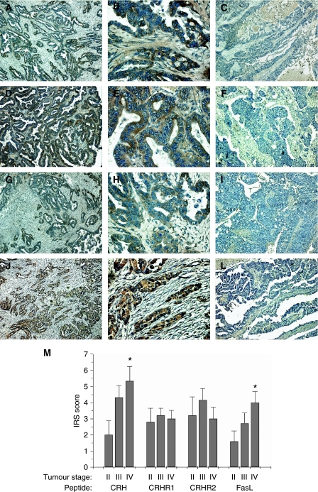
Immunohistochemical expression of CRH, CRHR1, CRHR2 and FasL in ovarian cancer tissue. (A–L) Representative photos of cases, which expressed or did not express the investigated peptides. CRH was immunolocalised in tumour cells in 68.1% of the cases examined (A, B), whereas the rest (31.9%) did not express the peptide (C). CRHR1 was expressed in 70.2% of cases (D, E) and absent from 29.8% (F). CRHR2 was expressed in 63.8% (G, H) of cases and absent from 36.2% (I). Finally, 63.8% of the cases were positive for FasL (J, K) and 36.2% were negative (L). Lens × 10 (A, C, D, F, G, I, J, L), × 40 (B, E, H, K). (M) Staining intensity was determined by the semiquantitative immunohistochemical IRS. There was a statistically significant increase in CRH and FasL expression between stage II and stage IV tumours. Data represent mean±s.e. (*P<0.05).

