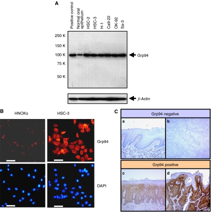
Figure 2.
Representative results of expression of Grp94 protein in oral squamous cell carcinomas (OSCC)-derived cell lines. (A) Western blot analysis of Grp94 protein in OSCC-derived cell lines and normal oral epithelium. All OSCC-derived cell line extracts exhibit a single band for Grp94 protein expression at high levels. In contrast, normal oral epithelium shows a low level of Grp94 protein expression. (B) Immunocytochemical analysis shows strong immunoreactivity of Grp94 in an OSCC-derived cell line (HSC-3) compared with human normal oral keratinocytes (HNOKs). DAPI staining was used to stain DNA. Bar, 100 μm. (C) Immunohistochemical staining of Grp94 in normal tissue, oral premalignant lesion (OPL), and primary OSCC. (a) Normal oral tissue exhibits negative Grp94 protein expression. (b) Grp94-negative case of OSCC. (c) Grp94-positive case of OPL. The immunoreaction is enhanced in the spinous layer. (d) Grp94-positive case of OSCC. Strong positive immunoreactivity for Grp94 is detected in the cytoplasm.

