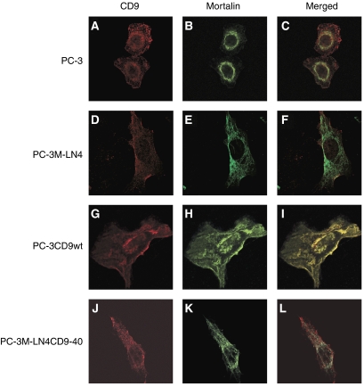
Figure 5.
Immunofluorescence analysis of cluster-of-differentiation antigen 9 (CD9) and mortalin in prostate cancer cells. Confocal laser scanning micrographs show staining for CD9 (red signals: A, D, G and J) and mortalin antigens (green signals: B, E, H and K). Overlap of signals in merged images revealed colocalisation of CD9 and mortalin in cells undergoing mitotic catastrophe (yellow staining in panel I). No protein colocalisation is detected in the other images (C, F and L).

