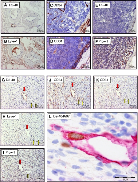
Figure 1.
Overview of the immunohistochemical stains used on serial sections of a metastatically uninvolved (perinodal fat: A–B; LN: C–F) and involved LN (G–L). In uninvolved LNs, high endothelial venules (*) show CD34 (C) and CD31 (D) reactivity. Littoral cells (+) are positive for CD31 (D) and Prox-1 (F). In metastatically involved LNs, podoplanin-positive LVs (red arrow) are demonstrated (G). Blood vessels (yellow arrow) are podoplanin negative (G). The LVs show CD34 (J), CD31 (K) and Prox-1 (I) reactivity. Although in perinodal fat (B) LVs are strongly positive for LYVE-1, the LVs in metastatically involved LNs are only faintly positive for LYVE-1 (H). Furthermore, a proliferating lymphatic endothelial cell is shown at the border of a LN metastasis (L) (LN: lymph node; LV: lymph vessel).

