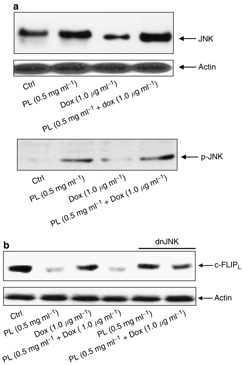
Figure 4.
JNK activation and c-FLIPL expression. (A) After treatment with PL, Dox or PL plus Dox, cells were harvested and lysates were prepared. The protein expression level of JNK or the presence of the phosphorylated form of JNK was determined by Western blot using the corresponding antibodies. Equal loading of total proteins in each sample was verified by β-actin. (B) c-FLIPL expression upon treatment with PL, Dox, or PL plus Dox. The cells with or without addition of a dn-JNK were treated with PL, Dox, or PL plus Dox. Subsequently, lysates were prepared to analyse the expression level of c-FLIPL. The percentages of the cells with fragmented DNA were determined by flow cytometry. The error bars represent the s.d. over five independent experiments.

