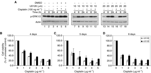
Figure 3.
Effect of MEK1/2 inactivation on cisplatin sensitivity in GH cells. GH cells were treated with two doses (10 and 20 μM) of an MEK1/2 inhibitor, U0126, for indicated time points. Western blotting and MTT assay were performed. (A) Western blotting analysis of p-ERK1/2 expression before (lanes 1–3) and after exposure to U0126 alone (lanes 7–9; 13–15) and in combination with cisplatin (lanes 10–12; 16–18). The cisplatin- and solvent-treated cells (lanes 4–6) were also tested as an internal control. Note that the expression of p-ERK was lower after exposure to both concentrations of U0126 compared with solvent control in response to cisplatin. (B–D) Cell viability after exposure to five concentrations of cisplatin for 4 days (B), 5 days (C) and 6 days (D) in the presence of 20 μM U0126 (filled columns) and absence of U0126 (open columns). Note that after exposure to both cisplatin and U0126, cell viability was higher than that treated with cisplatin alone. Results represented means of three independent experiments and error bars indicated standard deviation.

