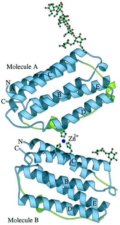
Figure 2.
Schematic representation of the crystallographic dimer of huIFN-β. The modeled portion of the carbohydrates and part of the zinc-binding site are also shown. The sphere corresponds to the zinc ion. Helices and N and C termini are labeled. The AB loop is colored green. The figure was made with the program molscript (44).

