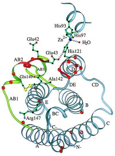
Figure 4.
Ribbon diagram of huIFN-β Cα backbone with side chains of residues known to be important for activity. The yellow spheres represent the sulfur atoms of the disulfide bridge. The ribbon is colored red at positions of the alanine-scanning mutagenesis cited in ref. 5. Orange spheres correspond to Cα atoms of residues homologous to those of IFN-α that are important for activity. Part of the zinc-binding site is also shown. The figure was made with the program molscript (44).

