
Figure 3.
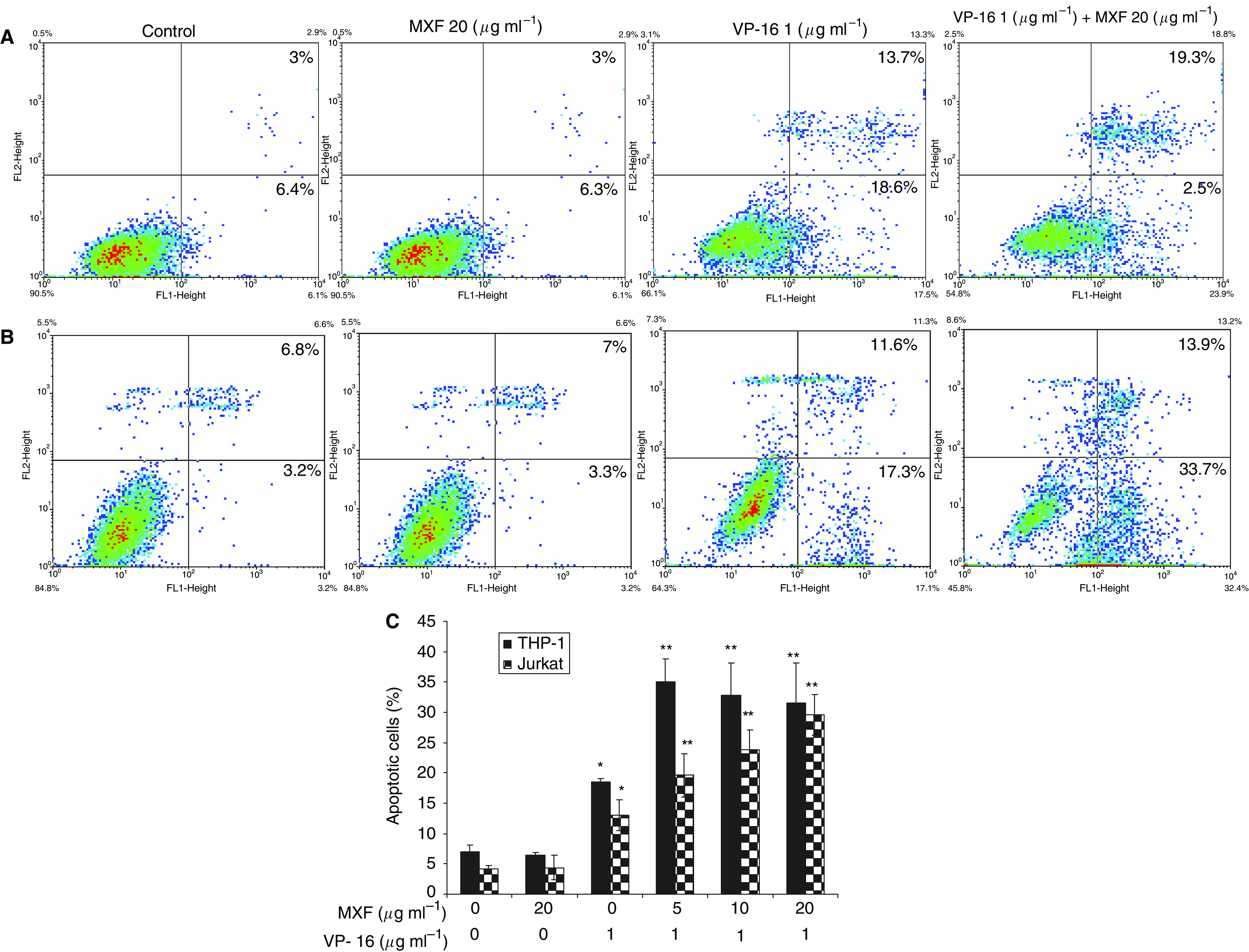
Moxifloxacin enhances apoptosis induced by VP-16. THP-1 (A and C) and Jurkat cells (B and C) were incubated for 24 h with VP-16 and MXF as indicated and flow cytometric analysis was performed by binding of annexin V and uptake of PI. A representative experiment is shown in (A and B). Results (mean±s.e.) of two independent experiments are shown in (C). The percentage of annexin V-positive, PI-negative cells is indicated in the lower right quadrangle and of annexin V-positive, PI-positive cells in the upper right quadrangle. The X-axis shows log annexin V fluorescence intensity and the Y-axis shows PI fluorescence intensity. *P<0.04 for cells treated with VP-16+MXF vs VP-16 alone.