Figure 4.
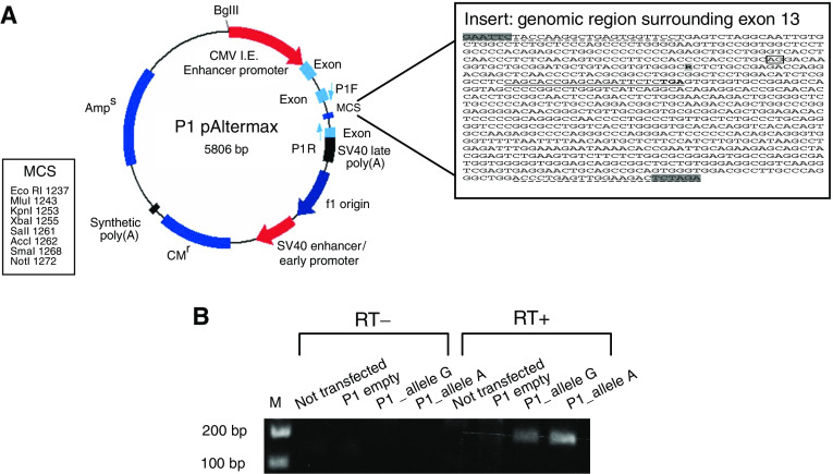
Splicing analysis for TIMM44 exon 13 variant. (A) P1 pAltermax plasmid structure, showing the two synthetic exons interrupetd by an intronic sequence carrying the multiple cloning site. Abbreviation: P1-F and P1-R primers forward and reverse specific for the synthetic exons of the vector. In the box it is shown the genomic sequence including exon 13 inserted in the P1 vector. In bold it is shown the change G1276A; underlined it is shown the sequence of the reverse primer specific for human TIMM44 encompassing the TGA stop codon; underlined and hatched it is shown the sequence of primers used for cloning, with the restriction sites shaded in grey. (B) RT–PCR results using the P1-F primer and the human primer specific for TIMM44 on the cDNA from COS7 cells not transfected (lanes 1 and 5) transfected with the empty P1 vector (lanes 2 and 6), allele G-containing vector (lanes 3 and 7), allele A-containing vector (lanes 4 and 8). Lanes 1–4: no superscript in the cDNA reaction mix; lanes 5–8 superscript present in the cDNA reaction mix. The PCR products are correctly visible only in the RT+ lanes of cells transfected with the vectors carrying the two alleles (lanes 7 and 8).

