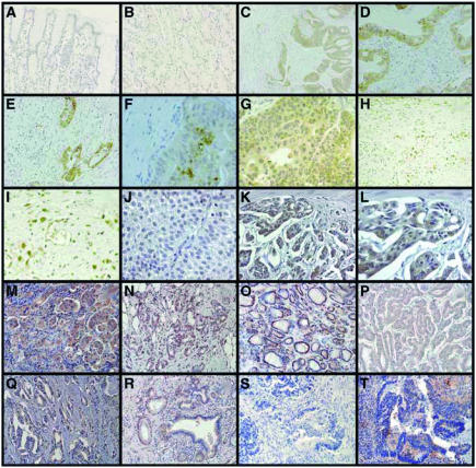
Figure 2.
Expression of TKTL1 in normal and carcinoma tissues. Specimens of a gastric carcinoma (C–G) and corresponding normal tissue (A, B); (A, B) no expression of TKTL1 in normal tissue. (C–G) Strong cytoplasmic expression in tumour tissue, but no expression in the surrounding stroma cells. Note the elevated expression within the inner region of the tumour (F). (H, I) Nuclear TKTL1 expression in a poorly differentiated gastric carcinoma. (J) No expression of TKTL1 in a superficial, Ta bladder carcinoma. (K, L) Strong TKTL1 cytoplasmic expression in an invasive, poorly differentiated bladder carcinoma. Strong TKTL1 upregulation in carcinomas of the lung (non-small-cell lung carcinomas; M), breast (N), thyroid (follicular thyroid carcinoma (O), papillary thyroid carcinoma (P)), prostate (Q), and pancreas (R). No expression of TKTL1 in a noninvasive colon carcinoma (S), and strong expression in an invasive colon carcinoma (T). Anti-TKTL1 was revealed by diaminobenzidine tetrahydrochloride (DAB; brown staining) (A–L) and 3-amino-9-ethylcarbazole (AEC; red staining) (M–T).

