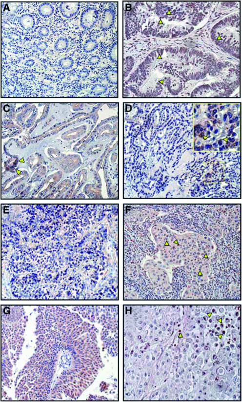
Figure 3.
Upregulation of phosphorylated Akt (p-AKT) in epithelial tumours. Immunohistochemical analysis of p-AKT on paraffin-embedded sections from normal colon as negative control (A), colon cancer (B), papillary (PTC) (C), follicular (FTC) (D), undifferentiated thyroid carcinoma (UTC) (E), non-small-cell lung cancer (NSCLC) (F), bladder cancer (G), and prostate cancer (H) (red staining). All the different types of cancer examined showed strong staining for p-Akt (cytoplasmic, nuclear, or both cytoplasmic and nuclear) while normal tissues showed no or very weak staining (A). A mainly nuclear localisation of p-Akt was been detected in colon, lung, and prostate carcinomas (B, F, H; yellow arrowheads). In all the histological variants of thyroid and bladder cancers, strong cytoplasmic staining was detectable (C–E, G) and only few nuclei in PTC and FTC samples were positive for p-Akt (C, D; yellow arrowheads).

