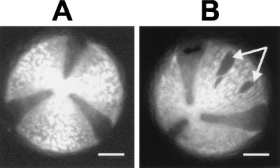
Figure 2.
Permeabilization of tobacco pollen grains. (A) Autofluorescence micrograph of tobacco pollen prior to permeabilization. (B) Pollen after permeabilization. The arrows indicate holes within the exine coat of the pollen. The dark sectors between the bright plates are not holes but are simply less fluorescent. (Bars = 10 μm.)

