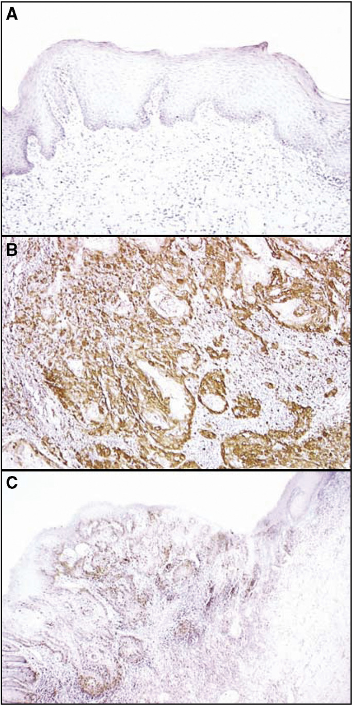
Figure 2.
Immunohistochemical staining of stathmin in normal and primary OSCCs. (A) Normal oral tissue shows no stathmin protein expression. Original magnification, × 200. (B) Stathmin-positive case of OSCC. Strong positive immunoreaction for stathmin is detected in the cytoplasm. Original magnification, × 200. (C) The border between normal epithelium (right side) and the dysplastic lesion (left side) is seen. While no stathmin expression is detected in normal epithelial cellular cytoplasm, strong stathmin protein expression is evident in the lesion. Original magnification, × 100.

