
Figure 2.
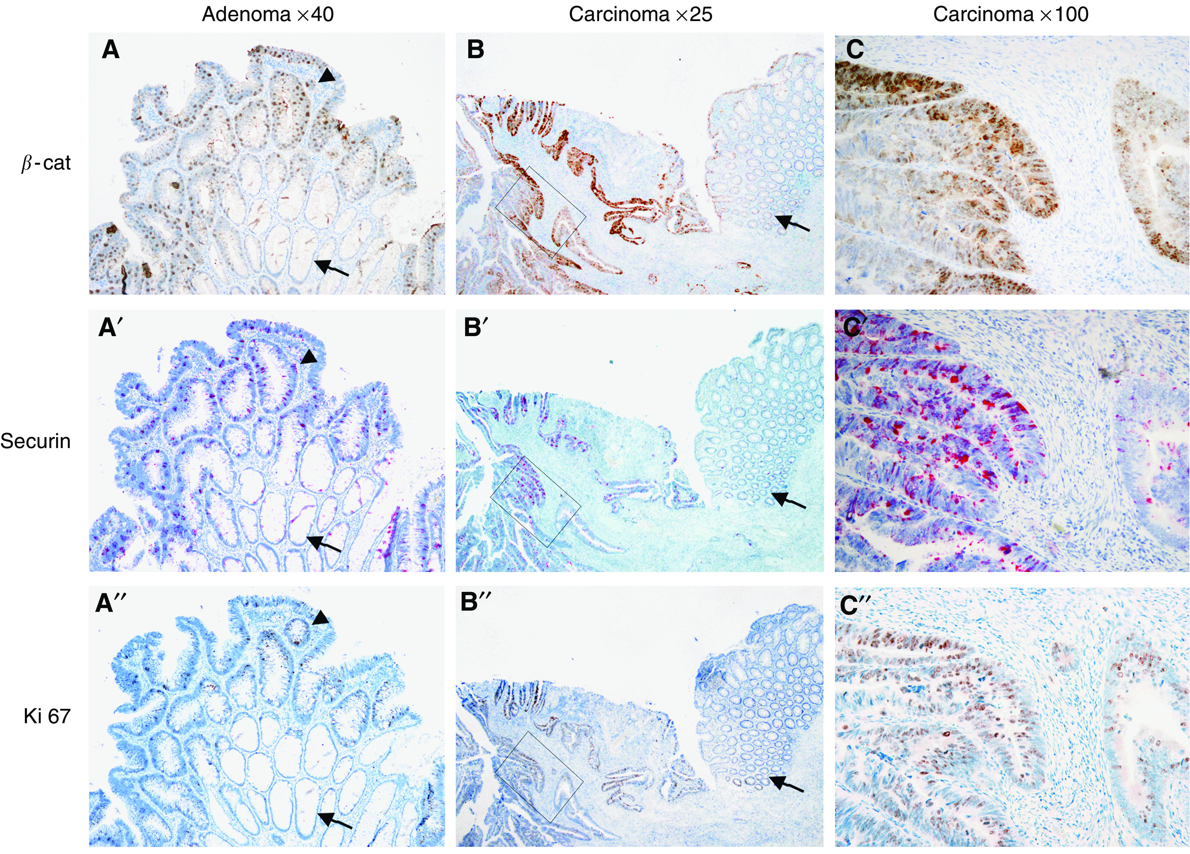
Correlated overexpression of nuclear β-catenin, securin and Ki-67 in colorectal adenoma and carcinoma. Immunohistochemical detection of β-catenin (first row), securin (second row) and Ki-67 (third row) using serial sections. Tissue sections show adenoma (a, a′, a′′), carcinoma (c, c′, c′′) and the normal mucosa (arrows in a, a′, a″ and b, b′, b′′) of the same case. Note that securin is already overexpressed in the adenoma tissue (arrowhead in a′) and only weakly expressed in the underlying normal colon mucosa (arrow in a′). A substantial overexpression of securin, Ki-67 and nuclear β-catenin can be seen in the carcinoma part (b, b′, b′′ and c, c′, c′′; the rectangle in b indicates the magnified area shown in c). Concordant with securin function in mitosis, the proliferation marker Ki-67 displayed the same expression pattern as securin. Nuclear accumulation of β-catenin in many of these tumour cells may suggest a function of this transcriptional activator protein for securin expression. Magnifications are indicated at the top of the figure.