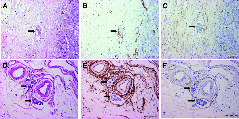
Figure 1.
Overview of the histological and immunohistochemical stainings on consecutive slides, used to differentiate between BVI (upper row: A, B and C) and LVI (lower row: D, E and F). Tumour cell emboli are indicated with black arrows. A and D: HE staining showing the presence of vascular invasion. B and E: On CD34 staining, both blood (C) and lymph (F) vessel endothelium stain positive. Furthermore, normal breast stromal cells are also CD34 positive (E). E and F: On D2-40 staining, the endothelium of vessels with BVI (B) and LVI (E) are respectively negative and positive. Desmoplastic stromal cells are also D2-40 positive (C). (BVI=blood vessel invasion, LVI=lymph vessel invasion).

