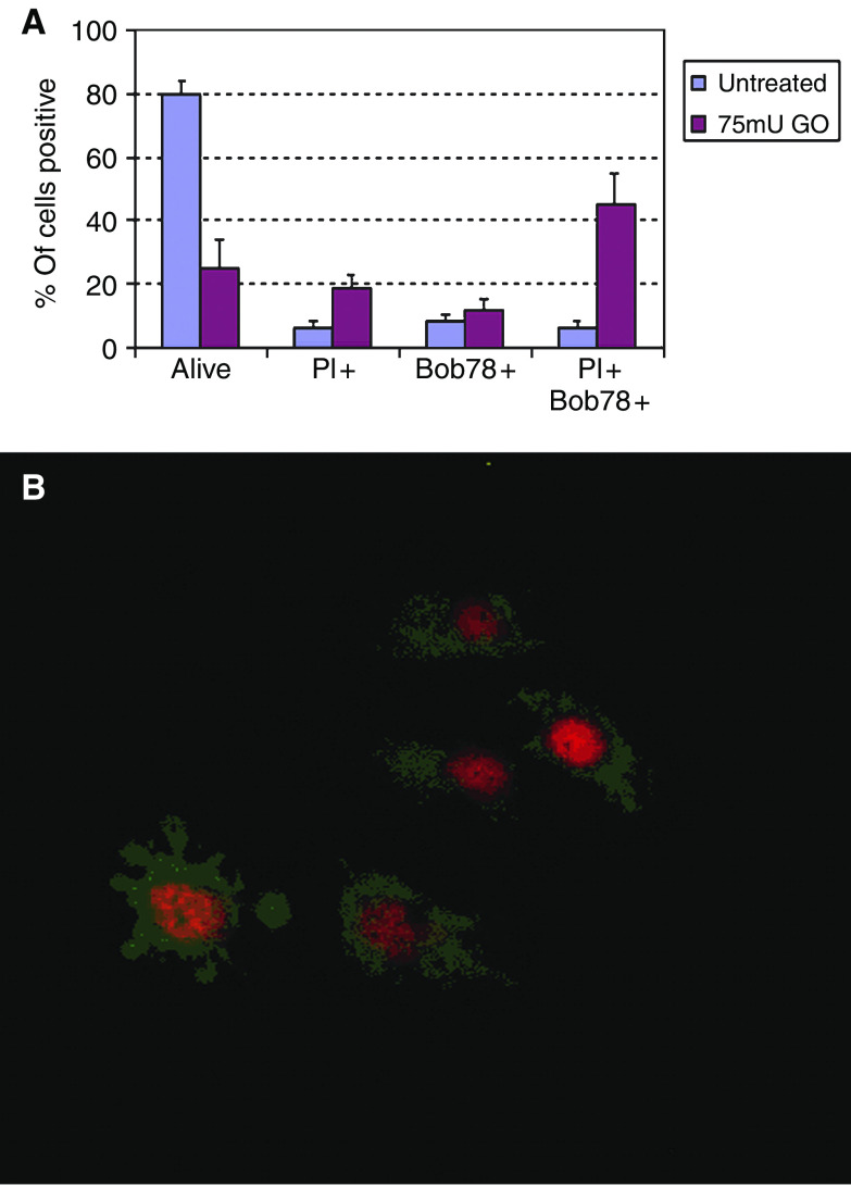
Figure 7.
Effect of glucose oxidase on BOB78 staining of HuH7 cells. (A) Flow cytometry. Following glucose oxidase treatment, cells were harvested, washed and co-stained with BOB78 antibody at 1/100 in PBS and propidium iodide. Treatment with glucose oxidase resulted in a decrease in nonstaining cells and an increase in both BOB78 only and double BOB78 and PI staining. (B) Immunocytochemistry. Cells were seeded at 2 × 10⁴ per well in eight-well chambered slides, incubated overnight and treated for 2 h with 75 mU ml−1 glucose oxidase. The medium was then harvested and cytospun on to charged slides at 300 g for 3 min. Cytospins were stained with BOB78 (1/100 in PBS) and propidium iodide and visualised using fluorescence microscopy. Cytospun cells again demonstrated predominantly necrotic morphology. A few cells with apoptotic features were seen (left), but again almost all of these were PI permeable. The pattern of BOB78 staining was predominantly cytoplasmic in necrotic cells and showed some redistribution to the plasma membrane in apoptosis, consistent with our previous findings.

