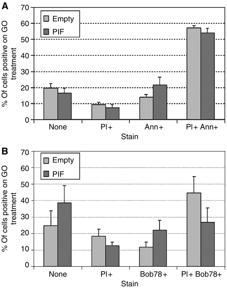
Figure 9.
Effect of transfection with wild-type PIF/dermcidin on flow-cytometric staining patterns of glucose oxidase-treated cells. (A) Annexin V. Cells were seeded at 1 × 10⁶ per well, treated with glucose oxidase, harvested, and stained with annexin V and propidium iodide as before. Proteolysis-inducing factor transfection resulted in a change in annexinV only staining from 15 to 22%, PI only staining from 9 to 8%, double staining from 57 to 54% and no staining from 19 to 16%. (B) BOB78. Cells were seeded at 1 × 10⁶ per well, treated with glucose oxidase, harvested, and stained with BOB78 (1/100 in PBS) and propidium iodide as before. The proportion of cells staining with BOB78 only increased from 12 to 22% on PIF transfection. BOB78 and PI double staining decreased from 45 to 27%. Nonstaining cells increased from 25 to 39% and PI only staining changed from 19 to 13%.

