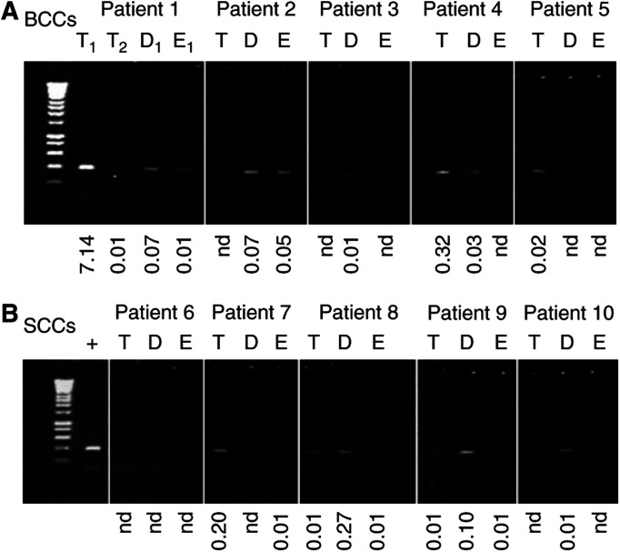
Figure 3.
Real-time PCR quantification and standard PCR amplification of the 3895 bp deletion in tumours. Ethidium bromide stained agarose gel showing the incidence of the 3895 bp deletion in tumour (T) and histologically normal perilesional dermis (D) and epidermis (E) from both BCCs (A) and SCCs (B) using the standard PCR assay. There are two different tumour samples (T1 and T2) from patient 1 (A); the associated dermis and epidermis are taken perilesional to T1. Below each lane is shown the level of the 3895 bp deletion illustrated as a percentage in each sample as quantified by real-time taqman PCR. Those samples marked with nd are determined to be effectively zero or ‘not detectable’ as the Ct of the real-time PCR was >36, which is the level observed in the no template control. Lane 1 in all panels=molecular weight markers (Hyperladder IV – range 1000–100 bp, Bioline Ltd, London UK). The positive (+) control in (B) is the tumour DNA from which the PCR product was cloned and sequenced to produce the template for real-time PCR. The same amount of template DNA was added to each PCR reaction. It worth noting that the greater sensitivity of the real-time PCR assay compared to the standard PCR assay is shown by the fact that detectable levels of the deletion are observed with the real-time assay even though there are no detectable PCR amplicons on the ethidium stained gel.

