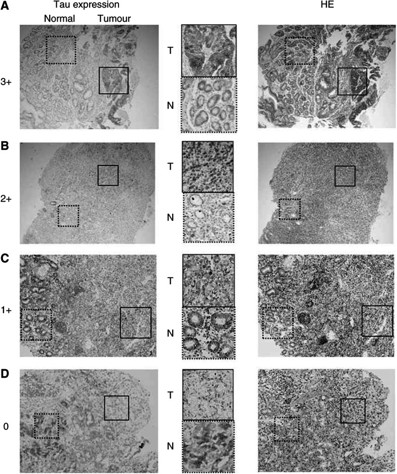
Figure 1.
Representative Tau expression pattern in gastric cancer cases for the IHC score (3+, 2+, 1+ and 0), whereas the right-hand side indicates the haematoxylin eosin staining (× 40 magnification). (A) 3+: There is strong granular staining in the cytoplasm of cancer cells obtained from biopsy specimens, moderately differentiated adenocarcinoma. This case was inoperable owing to the pleural effusions and the mediastinum lymph node metastases; a Paclitaxel nonresponsive case (PD). (B) 2+: Both nuclei and cytoplasm of the cancer cells are strongly positive. Normal mucosal glands are also positive. This case originated from a resected specimen of signet ring cell carcinoma after distal gastrectomy. A noncurative or a palliative operation was performed because of the dissemination of cancer cells into the retroperitoneum; a Paclitaxel nonresponsive case (PD). (C) 1+: A signet ring cell carcinoma case with positive tau expression is observed both in normal and tumour tissues. However, normal cells have a stronger signal than those of the tumorous tissue. No remarkable change was observed after Paclitaxel administration (NC). (D) 0: While the normal gastric glands (right) are strongly stained, the signet-ring type cancer cells (left) are completely negative. This case was partially responsive case (PR).

