
FIGURE 1. Sbh1p and Sbh2p participate in protein import into the ER in vivo.
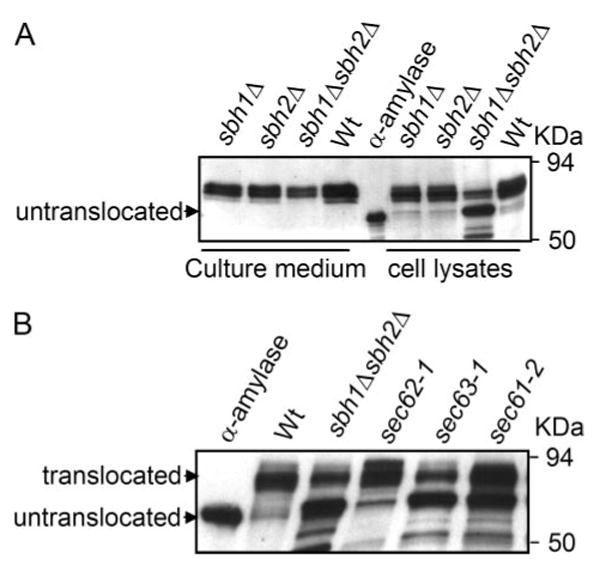
A, Western blot analysis of secreted and intracellular α-amylase. sbh1Δ (H3386), sbh2Δ (H3387), and sbh1Δ sbh2Δ (H3388) cells expressing α-amylase were grown at the permissive temperature and shifted to 37.5 °C for 9 h. Proteins present in the culture medium and cell lysate were resolved by SDS-PAGE, transferred to nitrocellulose membrane, and blots probed with antibodies against α-amylase. Untranslocated indicates the position of purified bacterial α-amylase. B, Western blot analysis of intracellular bacterial α-amylase in WT (H304), sbh1Δ sbh2Δ (H3232), sec62-1 (H1109), sec63-1 (H1107), and sec61-2 (H956) cells. Cells transformed with YEpαa6 were grown at 37.5 °C for 1 h, collected, and processed for SDS-PAGE and Western blotting with anti-α-amylase antibodies. α-Amylase indicates the lane loaded with purified bacterial α-amylase.