
FIGURE 2. Effect of Sbh1p and Sbh2p on in vitro protein import into the ER.
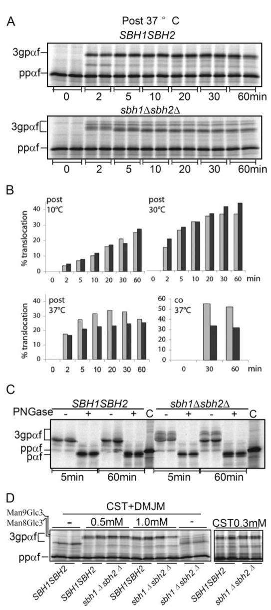
A, post-translational in vitro translocation of α-factor precursor (ppαf) into sbh1Δ sbh2Δ microsomes. Yeast microsomes (0.3 eq) were incubated with 300,000 cpm of ppαf translated in vitro in a yeast cell extract in the presence of ATP, and an ATP-regenerating system for the indicated periods of time at 37 °C. At the end of the incubation, samples were precipitated with trichloroacetic acid and analyzed by SDS-PAGE. B, quantitation of the post- and co-translational translocation of ppαf into sbh1Δ sbh2Δ microsomes in vitro at 10, 30, and 37 °C. Translocation was quantified and expressed as the percent of N-glycosylated protein (3gpαf) to total radiolabeled protein in each lane (3gpαf + ppαf). For co-translational import, 0.3 eq of membranes were used per 10 μl of in vitro translation/translocation reaction containing 2.3 μg of ppαf mRNA. Light gray, SBH1 SBH2; dark gray, sbh1Δ sbh2Δ. C, analysis of signal peptide cleavage of α-factor precursor in sbh1Δ sbh2Δ cells. In vitro translated ppαf was translocated into wild type and mutant membranes for 5 or 60 min, the membranes were lysed and glycosylated 3gpαf precipitated with concanavalin A-Sepharose. Precipitated 3gpαf was incubated with N-glycanase to remove N-linked glycans or mock-digested, and deglycosylated, translocated proteins were analyzed by SDS-PAGE on 18% 2 m urea gels, aliquots of in vitro translated ppαf containing the signal peptide were loaded as controls (C). D, effect of castanospermine (CST) and 1-deoxymannojirimycin (DMJM) on migration of 3gpαf. Membranes from sbh1Δ sbh2Δ and WT cells were preincubated with the indicated concentrations of castanospermine and/or 1-deoxymannojirimycin and during the translocation reactions followed by gel analysis. Positions of 3gpαf with Man9Glc3 and Man8Glc3 glycans are indicated on the left.