
FIGURE 3. Sbh1p and Sbh2p are dispensable for pΔgpαf ERAD.
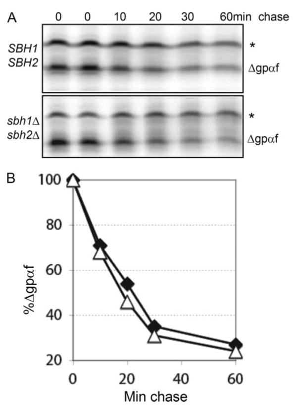
A, mutant α-factor precursor (pΔgpαf) was translocated into wild type or mutant microsomes, membranes were washed, and ERAD was initiated by adding yeast cytosol ATP and an ATP-regenerating system, and incubation at 30 °C. At the indicated time points, samples were precipitated with trichloroacetic acid and proteins analyzed by SDS-PAGE and phosphorimaging. The asterisk indicates untranslocated pΔgpαf associated with the cytosolic face of the microsomes. B, quantitation of Δgpαf on the gels shown in A. △, SBH1 SBH2; ♦, sbh1Δ sbh2Δ.