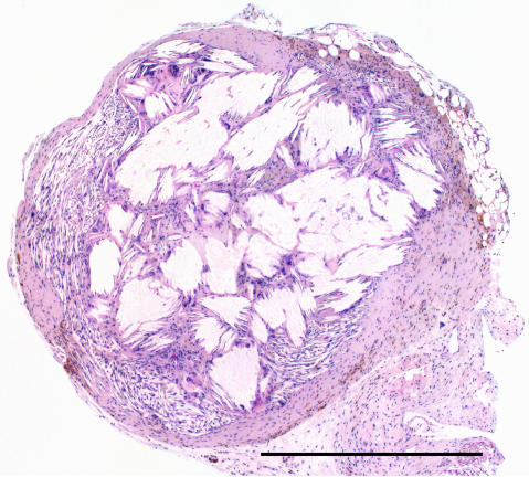
Figure 6.
Histopathological analysis of a small remnant of tumour tissue in an animal with CR 5 months after administration of 120 MBq [177Lu-DOTA0-Tyr3]-octreotate. The tumour residue consisted of a brownish nodule (2 mm in diameter). In the periphery, only a thin rim of fibroblasts was found. The dominant part of the nodule contained crystalline structures surrounded by inflammatory cells including macrophages and giant cells. Scale bar equals 1 mm.

