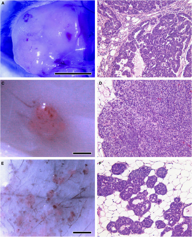
Figure 1.
Digital image capture of PpIX-stimulated red fluorescence in tumours of three mammary tumour-bearing transgenic strains 1 h following in vivo administration of 200 mg kg−1 ALA, with corresponding H&E stained histological sections. (A, B) Red fluorescence in a papillary adenocarcinoma of a WapTag1 female (77 weeks) (20 ×). (C, D) HRAS male mammary tumour (11 weeks) showing prominent fluorescence and solid carcinoma histology (20 ×); (E, F) Fluorescent multifocal tumours of a PyVT female (5 weeks) with solid carcinoma histology (20 ×). Scale bar represents 1 mm.

