Figure 1.
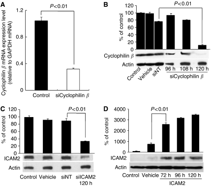
Effect of siRNAs. (A) Expression of cyclophilin β mRNA in non-transfected HSC2 cells (control) and HSC2 cells transfected with siCyclophilin β (P<0.01, Student's t-test). (B) Western blot analysis of cyclophilin β protein (positive control) in HSC2. The cells were transfected with 100 nmol l−1 siNT and siCyclophilin β and analysed at 96, 108, and 120 h. Cyclophilin β and actin bands were scanned and quantitated as described under Materials and Methods. The values obtained from densitometric analysis of each cyclophilin β protein first were normalised to actin protein levels and then expressed as the percentage of the values of control. Cyclophilin β proteins were significantly inhibited (P<0.01, Student's t-test) in cells transfected with siCyclophilin β at 120 h. There is no change in cyclophilin β in cells transfected with vehicle and siNT negative controls siRNA. (C) Western blot analysis of ICAM2 protein in HSC2 cells transfected with vehicle, siNT, and siICAM2. The cells were transfected with 100 nmol l−1 siRNAs and analysed at 120 h. Intercellular adhesion molecule 2 and actin bands were scanned and quantitated as described under Materials and Methods. The values obtained from densitometric analysis of each ICAM2 protein first were normalised to actin protein levels and then expressed as the percentage of the values of control. The ICAM2 proteins were significantly inhibited (P<0.01, Student's t-test) in cells transfected with siICAM2 at 120 h. (D) Western blot analysis of ICAM2 in ICAM2-overexpressing HSC3 cells. The cells were examined 72, 96, and 120 h after transient transfection of expression vector encoding ICAM2 cDNA. Actin was used as a loading control. Intercellular adhesion molecule 2 and actin bands were scanned and quantitated as described under Materials and Methods. The values obtained from densitometric analysis of each ICAM2 protein first were normalised to actin protein levels and then expressed as the percentage of the values of control. The ICAM2 protein levels increased significantly (P<0.01, Student's t-test) in cells transfected with the expression vector of ICAM2 DNA at 72, 96, and 120 h.

