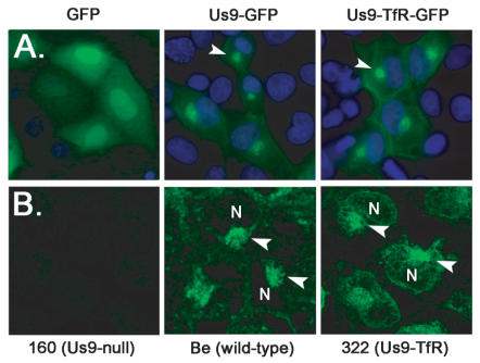
Figure 6. Localization of Us9-TfR in the absence and presence of infection.
(A) PK15 cells were transfected with plasmids expressing GFP, Us9-GFP, and Us9-TfR-GFP. Cells were fixed with 4% paraformaldehyde at 24 hours post-transfection, and the nuclei stained with Hoechst 33342 (blue). Direct fluorescence was visualized using an inverted epifluorescence microscope using the appropriate excitation and emission filters. The arrowheads highlight the perinuclear, steady-state accumulation of Us9. (B) PK15 cells were infected with PRV 160 (Us9-null), Becker (wild-type), or PRV 322 (Us9-TfR) for 6 hours. Cells were fixed and stained with Us9 antiserum, and visualized on a Leica SP5 confocal microscope. Arrowheads denote Us9 accumulations adjacent to the cell nucleus (N).

