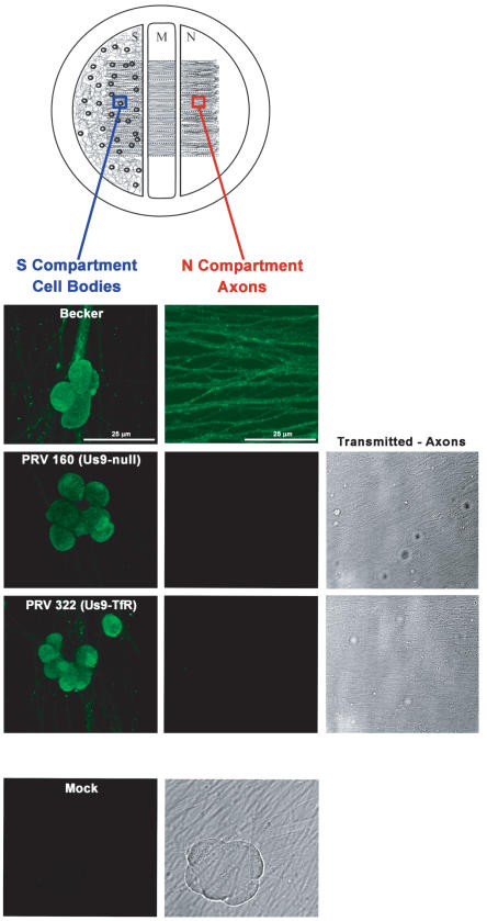
Figure 8. PRV 322 (Us9-TfR) is defective in axonal sorting of virus structural proteins.
Trichamber diagram illustrating the system used to visualize PRV antigens in the cell bodies and axons of infected neurons. The blue box illustrates the site within the S compartment were cell bodies were imaged. The red box indicates the site were axons were imaged in the N compartment. SCG neurons were plated in the S chamber and allowed to extend neurites into the N chamber (through the M compartment). The neurites were guided into the N compartment by a series of grooves. Two weeks post-plating, cell bodies in the S chamber were infected at a high MOI with Becker (wild-type), PRV 160 (Us9-null) or PRV 322 (Us9-TfR). At 16 h postinfection, samples were fixed and labeled with PRV-specific polyclonal antiserum (Rb134) that recognizes virus glycoprotein and virus capsid proteins. All infected cell bodies within the S compartment stained for viral structural proteins (first column). Mock-infected cells did not label with the Rb134 antibody. Becker-infected axons stained heavily for PRV antigen (second column), though axons from PRV 160 and PRV 322 infected cell bodies were devoid of viral glycoprotein and capsid proteins (though an extensive network of axons was visible by transmitted brightfield illumination).

