
Figure 3. Analysis of UPR in a cortical tuber dissected from a 3 year-old patient with tuberous sclerosis.
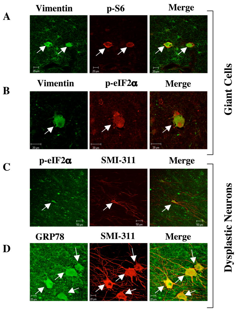
(A) Vimentin and phospho-S6 (ser235/236) staining of the tuber identify enlarged glial cells in which the mTORC1 pathway has been activated. Adjacent tissue contains many weakly-vimentin positive astroglia that do not contain phospho-S6 immunoreactivity. (B) Vimentin and phospho-eIF2α (ser51) staining in a tuber shows an enlarged cell that contains immunoreactivity for both glial marker vimentin and UPR marker phospho-eIF2α. (C) SMI-311 and phospho-eIF2α staining in a tuber shows an enlarged neuron, which contains immunoreactivity for the UPR marker phospho-eIF2α (ser51). Scale bar represents 50μm (D) SMI-311 and GRP78 staining in a tuber section shows several enlarged neurons that are immuno-reactive for the UPR marker GRP78. Scale bar represents 20μm.