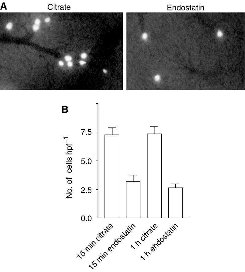
Figure 3.
(A) Intravital microscopy images recorded 15 min after injection of fluorescent tumour cells into the spleen of a control liver and a liver after 2 h rh-E pretreatment. (B) Number of fluorescent tumour cells that are present in the liver per hpf as measured by intravital microscopy 15 min and 1 h after intrasplenic injection. Recombinant human endostatin reduced the number of arrested tumour cells in the liver by 56% (P<0.001). No differences were observed within either treatment group between the two time points (controls 15 min vs 1 h P=0.93; endostatin 15 min vs 1 h P=0.39).

