Figure 4.
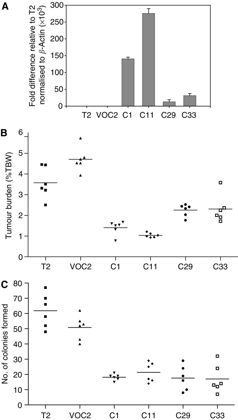
Expression of the Testisin gene suppresses tumorigenicity of Tera-2 cells in vivo and inhibits anchorage-dependent colony formation in vitro. (A) Testisin mRNA expression in transfected Tera-2 cell lines. Graph showing fold difference in Testisin mRNA expression levels relative to β-actin determined by quantitative real-time PCR: mRNA isolated from parental Tera-2 cells [T2], Tera-2 clones transfected with Testisin cDNA [C1, C11, C29, C33] or the control Tera-2 clone transfected with vector alone [VOC2]. (B) Testisin gene expression suppresses Tera-2 tumour growth in a murine in vivo orthotopic xenograft model of testicular tumorigenesis. Tumour burden (testis tumour weight as a percentage of the total mouse body weight) was calculated as described in the Materials and Methods and is displayed as a scattergram with the line centered on the mean of the values. C1, C11, C29, C33 vs VOC2, P=0.0022; Kruskal–Wallis nonparametric test. There was no significant difference between parental Tera-2 cells and the vector alone control, T2 vs VOC2, P=0.0877. (C) Testisin gene expression inhibits anchorage-dependent colony forming ability of Tera-2 cells in-vitro. Cells were plated at low density and colony formation monitored over 14 days with media changed every 4 days. C1, C11, C29, C33 vs VOC2, P=0.0022; Kruskal–Wallis nonparametric test. There was no significant difference between parental Tera-2 cells and the vector alone control, T2 vs VOC2, P=0.1320.

