Figure 5.
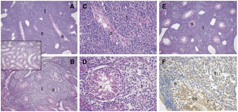
Photomicrographs of histological analyses of murine orthotopic testicular tumours. Tissue sections were stained with Mayers’ haematoxylin and eosin. (A, B) Examples of severely atrophied residual seminiferous tubules (s) present in testes containing testicular tumours (t) formed after 4 weeks following injection of the vector alone cell line control. (A) and (B) are × 100 and × 400 magnification respectively. (C) Representative residual seminiferous tubule with moderate atrophic changes present in testes 4 weeks following injection of C33 (× 100 magnification). (D, E) Examples of residual seminiferous tubules with mild atrophic changes after 4 weeks following injection of C11, at magnifications of × 100 and × 400, respectively. (F) Immunohistochemical staining for Testisin in a section containing C11 tumour at × 400 magnification. The monoclonal antibody (DD-P104 C37) reacts with human Testisin present in the tumour (t) as well as murine Testisin present in the round (r) and elongating spermatids (es) of the murine seminiferous tubules. The inset (× 100 original magnification) shows a contralateral testis with normal morphology.

