
Figure 1.
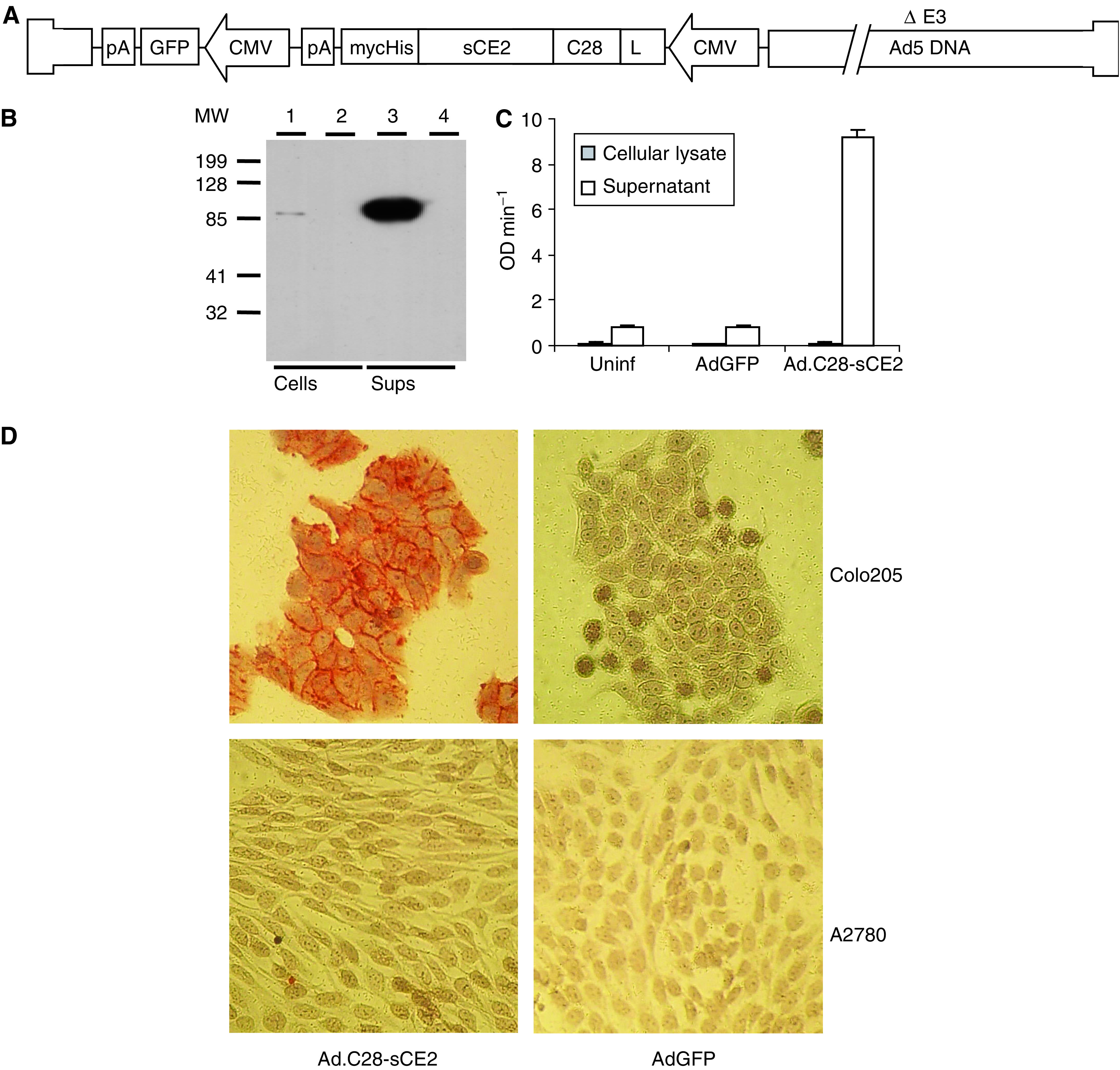
Schematic structure of the replication-deficient adenovirus Ad.C28-sCE2 and characterisation of Ad.C28-sCE2-transduced SW1398 cells by Western blot analysis, esterase activity assay and immunohistochemistry. (A) Schematic structure of the replication-deficient adenovirus Ad.C28-sCE2. The C28-sCE2 expression cassette includes the CMV promoter, an IgGκ leader sequence for secretion and a C-terminal myc- and His-tag for detection and purification. The adenovirus also contains the gene encoding GFP under the CMV promoter. (B) Western blot analysis of cellular lysates (lanes 1 and 2) and supernatants (lanes 3 and 4) of SW1398 cells transduced with Ad.C28-sCE2 (lanes 1 and 3) or AdGFP (lanes 2 and 4) at an MOI of 100. C28-sCE2 was detected using an antibody directed to the myc-tag. (C) CE activity in cellular lysates and supernatants of SW1398 cells transduced with Ad.C28-sCE2 or AdGFP at an MOI of 100. Cellular lysates or supernatants were incubated with 1 mM pNpAc and conversion was measured during 10 min. C28-sCE2 showed enzymatic activity and was efficiently secreted by transduced cells, since most of the activity was detected in the supernatant. (D) Binding of C28-sCE2 to the EpCAM-expressing cell line Colo205. Colo205 cells or the EpCAM-negative cell line A2780 were incubated with the supernatant of SW1398 cells transduced with Ad.C28-sCE2 or AdGFP at an MOI of 100. After washing, the cells were stained with anti-myc antibody to show binding of C28-sCE2. Only the EpCAM-expressing Colo205 cells incubated with supernatant of Ad.C28-sCE2-transduced SW1398 cells showed a positive membrane staining, indicating that the fusion protein had bound specifically to the Colo205 cells.