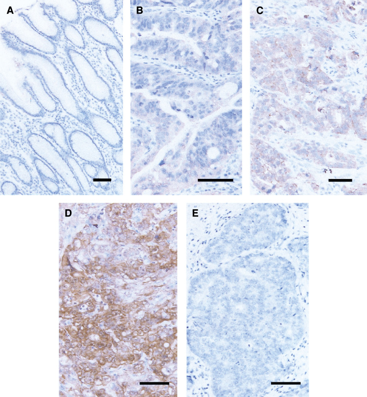
Figure 2.
Immunohistochemistry for maspin protein. (A) High power view of gastric normal foverolar epithelium (scale bar=100 μm). Immunoreactivity for maspin is negative. (B–D) High power view of maspin-positive gastric cancers (scale bar=100 μm). Subcellular localisation of maspin protein is observed in cytoplasm and membrane (B–D). (B) (Case No. 24) and (C) (Case No. 32) are moderately differentiated tubular adenocarcinomas, and (D) (Case No. 14) is a poorly differentiated adenocarcinoma, solid type. (E) High power view of maspin-negative gastric cancers (Case No. 10; scale bar=100 μm).

