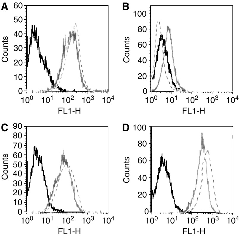
Figure 1.
FACS analysis of (A) A431 adenocarcinoma cell line (amplified EGFR) and (B) parental and (C) transfected U87MG glioma cell lines stably expressing wt (U87MG.wtEGFR) or (D) mutant EGFR (U87MG.de2-7). Cells were incubated with mAb806 (–), ch806 (- -) followed by Alexa488-labelled anti-mouse Ig. The plots represent fluorescence intensity on the abscissa and cell number per fluorescence channel on the ordinate. The negative control (irrelevant antibody) fluorescence is plotted on each panel (black line).

